Brain · Tumours
Brain Tumour Evaluation & Surgery in Faridabad
Clear guidance from the first scan to the final plan, so you understand whether to watch, biopsy, operate or treat with radiotherapy.
हर ब्रेन ट्यूमर कैंसर नहीं होता और हर ट्यूमर में ऑपरेशन की ज़रूरत भी नहीं होती। MRI, बायोप्सी और ज़रूरत पड़ने पर सर्जरी के ज़रिए हर मरीज़ के लिए अलग इलाज योजना बनाई जाती है।
Understanding Brain Tumour Surgery
A brain tumour is an abnormal growth of cells inside the skull. The word is frightening, but it covers a very wide range of conditions. Some tumours are benign: slow-growing and not cancerous, such as most meningiomas. Others are malignant, like a high-grade glioma, which grows faster into nearby brain. Tumours may start in the brain itself (primary) or spread there from cancer elsewhere (metastasis).
Not every brain tumour needs an operation. A small tumour found by chance may only need watching with repeat MRI, and some respond to medicines or radiotherapy. When surgery is advised, the aim may be complete removal, safe partial removal to relieve pressure, or a biopsy to identify the tumour exactly so the right treatment can follow.
For families in Faridabad, Ballabhgarh and Delhi NCR, the first step is a calm review of scans and symptoms. At Sarvodaya Hospital, Sector 8, care is planned with radiology, pathology and oncology colleagues so each decision fits the individual patient.
Common symptoms
Symptoms vary from person to person. Having one of these does not mean you have this condition.
New headaches that are worse in the morning or on coughing, sometimes with vomiting.
A first fit in an adult always needs a brain scan; it can be the first sign of a tumour.
Slowly increasing weakness of an arm, leg or one side of the face.
Blurred or double vision, loss of side vision, or hearing loss in one ear.
Trouble finding words, confusion or forgetfulness noticed by family.
Possible causes & risk factors
For most primary brain tumours no definite cause is found; nothing the patient did caused it.
Radiotherapy to the head, especially in childhood, slightly raises the risk.
Rare conditions such as neurofibromatosis increase the chance of certain tumours.
Lung, breast, kidney and skin cancers can spread to the brain.
When to seek help
Book a routine consultation
- Headaches that are new or steadily worsening over weeks
- A tumour found incidentally on an MRI done for another reason
- Slowly increasing weakness, numbness or unsteadiness
- A second opinion after being told surgery is needed
Seek emergency care
- A first seizure, or a fit lasting more than five minutes
- Sudden severe headache with repeated vomiting or drowsiness
- Sudden weakness, loss of speech or loss of vision
- Confusion or difficulty waking the person: go to the nearest emergency department or call 112
How it is diagnosed
The key scan; shows the tumour's size, location and relation to important brain areas.
Perfusion, spectroscopy or tract mapping can help predict tumour type and plan a safe route.
Tissue examined under the microscope, with molecular tests, gives the final diagnosis.
Available treatment options
Treatment is chosen after examining you and studying your scans. Many people do well without an operation.
Small, benign-looking tumours without symptoms are often monitored with planned scans. Many never need surgery.
Steroids may ease brain swelling (oedema) and anti-seizure medicines control fits.
A navigation-guided needle takes a small sample through a small opening, useful for deep tumours.
A window of skull bone is lifted temporarily so the tumour can be removed under the microscope as far as is safe.
Selected tumours near the ventricles or skull base may be reached through smaller openings with neuroendoscopy.
Planned by the oncology team after biopsy or surgery, depending on tumour type and grade.
When neurosurgery may be recommended
- The tumour causes symptoms by pressing on the brain
- Follow-up MRI shows steady growth
- Tissue is needed to plan radiotherapy or chemotherapy
- There is risk of blocked brain fluid (hydrocephalus) or brain shift
- The tumour can be reached with acceptable risk to speech, movement and vision
Procedure overview
A simplified outline. The exact steps depend on your scans and the approach chosen.
- PlanningMRI is loaded into the navigation system and a route is planned around key brain areas.
- AnaesthesiaMost operations are done under general anaesthesia with the head held still.
- Removal or samplingThe tumour is removed under magnification, or a biopsy sample is taken.
- Close monitoringThe bone is replaced and the patient is watched in the neuro ICU, usually overnight or longer.
Potential benefits, risks & limitations
Potential benefits
- May relieve headache, seizures or weakness caused by pressure
- Gives a definite diagnosis to guide further treatment
- Reducing tumour bulk may help radiotherapy and chemotherapy work better
- Can lower the risk of sudden deterioration from swelling
Possible risks
- Bleeding or infection
- New or worsened weakness, speech or vision problems, sometimes temporary
- Seizures or brain swelling after surgery
- Tumour regrowth needing further treatment
Surgery cannot always remove every tumour cell, especially where a tumour grows into healthy brain. For malignant tumours, surgery is one part of a longer plan rather than a complete answer on its own.
Recovery & follow-up
Many patients walk within a day or two and go home within several days, though this varies with the tumour, age and general condition.
Some patients benefit from physiotherapy or speech therapy. The final pathology report decides whether radiotherapy, chemotherapy or simply follow-up MRI comes next. Our guide on what happens during brain tumour evaluation explains each stage.
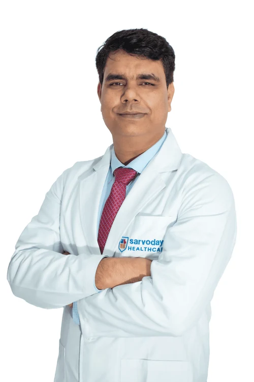
Why consult Dr. Naresh Panwar
- MCh (Neurosurgery), SMS Medical College, Jaipur, with a Fellowship in Neuroendoscopy
- Senior Consultant & Head Unit (II) – Neurosurgery, Sarvodaya Hospital, Sector 8, Faridabad
- Published case imaging includes navigated (frameless) brain biopsy and meningioma cases
- Brain tumour surgery and skull base surgery are listed specialisations on his hospital profile
Questions to ask your surgeon
Write these down and bring them to your appointment.
- What type of tumour do you think this is?
- Is watching with scans safe in my case?
- Is the aim complete removal, partial removal or biopsy?
- Which functions could be affected, and how will you protect them?
- Will I need radiotherapy or chemotherapy afterwards?
Frequently asked questions
Is every brain tumour cancer?
No. Many brain tumours, such as most meningiomas and pituitary adenomas, are benign. They can still cause problems by pressing on the brain. Only tissue testing confirms the exact type.
What is a navigated brain biopsy?
A thin needle is guided to the tumour by computer navigation linked to your MRI, taking a small sample through a small skull opening. It is often chosen for deep tumours or to confirm the diagnosis before further treatment.
Brain tumour ka ilaaj kya bina operation ke ho sakta hai?
Kuch cases mein haan. Chhote, dheere badhne wale tumours ko kai baar sirf MRI se monitor kiya jaata hai, aur kuch dawai ya radiotherapy se control hote hain. Sahi raasta reports aur jaanch ke baad hi tay hota hai.
I live in Ballabhgarh. Where can I consult for a brain tumour?
Consultations are at Sarvodaya Hospital, Sector 8, Faridabad, near Escorts Mujesar Metro Station (Violet Line). Call or WhatsApp +91 96608 88640 to confirm timings, and bring all MRI films.
How long does a brain tumour operation take?
It varies from under an hour for a biopsy to several hours for a complex tumour. Your surgeon can estimate after studying your scans.
Related care & reading
Appointments
Your brain and spine health deserve a carefully considered plan.
Request a consultation with Dr. Naresh Panwar at Sarvodaya Hospital, Sector 8, Faridabad. Bring your MRI or CT scans and reports, and the care team will help you prepare.