Brain · Blood Vessel Disorders
Cerebrovascular Neurosurgery in Faridabad
When a brain blood vessel is malformed, weakened or has bled, the right plan comes from careful imaging and a team that weighs every option, including watching.
AVM, कैवर्नोमा, एन्यूरिज़्म और ब्रेन हेमरेज जैसी दिमाग की नसों की समस्याओं में सही जांच और टीम-आधारित योजना ज़रूरी है। हर मरीज़ को सर्जरी की ज़रूरत नहीं होती — कई बार नियमित निगरानी ही सही रास्ता होता है।
Understanding Cerebrovascular Surgery
Cerebrovascular neurosurgery deals with problems in the blood vessels of the brain. Some are present from birth, such as an arteriovenous malformation (AVM), a tangle where arteries join veins directly, or a cavernoma, a small mulberry-like cluster of thin-walled vessels. Others develop during life, such as a bulge in an artery wall called an aneurysm, or narrowing of the carotid arteries in the neck.
The main worry is bleeding. A haemorrhagic stroke happens when a vessel inside the brain bursts, and many Faridabad families first hear the word AVM in an emergency room. Our brain haemorrhage treatment page covers that emergency; this page explains the vessel problem underneath and the longer-term plan.
Not every abnormal vessel needs an operation. Many small cavernomas and some unruptured aneurysms are simply watched with periodic scans. When treatment is advised, choices include open microsurgery, catheter-based endovascular treatment, focused radiation, or a combination. Dr. Naresh Panwar discusses these with neuro-interventional and critical-care colleagues so the plan fits the vessel and the patient.
Common symptoms
Symptoms vary from person to person. Having one of these does not mean you have this condition.
A thunderclap headache, often called the worst ever felt, can signal bleeding and needs emergency care.
A first fit in an adult is a common way AVMs and cavernomas come to light.
One-sided weakness, facial droop or numbness may follow a bleed or reduced blood flow.
Short-lived weakness, or vision loss in one eye, can point to carotid artery narrowing.
Many vessel abnormalities are found incidentally on an MRI done for another reason.
Possible causes & risk factors
AVMs and most cavernomas form before birth; nothing the patient did caused them.
Uncontrolled blood pressure weakens small brain arteries and is a major cause of haemorrhagic stroke.
Raises aneurysm risk and speeds up artery narrowing.
These drive the fatty build-up that narrows the carotid arteries.
When to seek help
Book a routine consultation
- An AVM, cavernoma or aneurysm reported on a scan done for something else
- A first seizure that has settled, with a vessel abnormality on MRI
- A relative with a known brain aneurysm, asking about screening
Seek emergency care
- Sudden, explosive headache, especially with vomiting or a stiff neck — go to the nearest emergency department or call 112
- Sudden facial droop, arm weakness or slurred speech — learn the stroke warning signs and call 112
- Fainting, confusion or drowsiness after a severe headache
- A seizure lasting more than five minutes
How it is diagnosed
The quickest emergency test; shows bleeding and outlines the major arteries.
Shows cavernomas, old bleeds and surrounding brain tissue in fine detail.
A catheter dye study, done by the neuro-interventional team, that maps blood flow precisely for planning.
A simple neck scan measuring carotid narrowing.
Available treatment options
Treatment is chosen after examining you and studying your scans. Many people do well without an operation.
Small, deep or never-bled lesions are often safest left alone, with regular imaging to catch change early.
Blood pressure control, anti-seizure medicines where needed and stopping smoking lower the chance of future problems.
Neuro-interventional specialists can treat some aneurysms and AVMs from inside the vessel via a catheter. Suitability is decided by the treating team.
Open surgery under a microscope can remove an AVM or cavernoma, or secure an aneurysm, when this is the safer route.
For some small, deep AVMs, targeted radiation slowly closes the vessels over time; where suitable, the team will explain referral.
When neurosurgery may be recommended
- A cavernoma or AVM that has bled and can be reached safely
- Seizures continuing despite medicines and linked to a removable lesion
- A large blood clot pressing on the brain after a haemorrhage
- Aneurysm shapes better suited to open repair than catheter treatment
Procedure overview
A simplified outline. The exact steps depend on your scans and the approach chosen.
- Joint case reviewScans are reviewed with neuro-interventional and critical-care colleagues to compare options.
- PreparationBlood tests, anaesthesia check-up and adjustment of blood thinners are planned.
- MicrosurgeryUnder general anaesthesia, a small skull window lets the surgeon work under magnification.
- Imaging and ICU careA scan or angiogram checks the result; early recovery is usually in the neuro-ICU.
Potential benefits, risks & limitations
Potential benefits
- May lower the risk of a future bleed from the treated vessel
- Can help seizure control when a cavernoma or AVM is the source
- Relieves pressure on the brain when a clot is removed
Possible risks
- Bleeding during or after surgery
- New weakness, speech or vision problems
- Seizures after the operation
- Infection or wound-healing problems
- Need for further treatment if part of the lesion remains
Treatment cannot reverse damage from a previous bleed, and some deep lesions are safer to watch than to operate on.
Recovery & follow-up
Recovery varies widely. Someone treated for an unruptured cavernoma may go home within days, while recovery from a major bleed may need weeks of rehabilitation.
Families travelling from Ballabhgarh, Palwal or Greater Faridabad are usually advised repeat imaging at intervals set by the team. Never stop seizure or blood pressure medicines on your own.
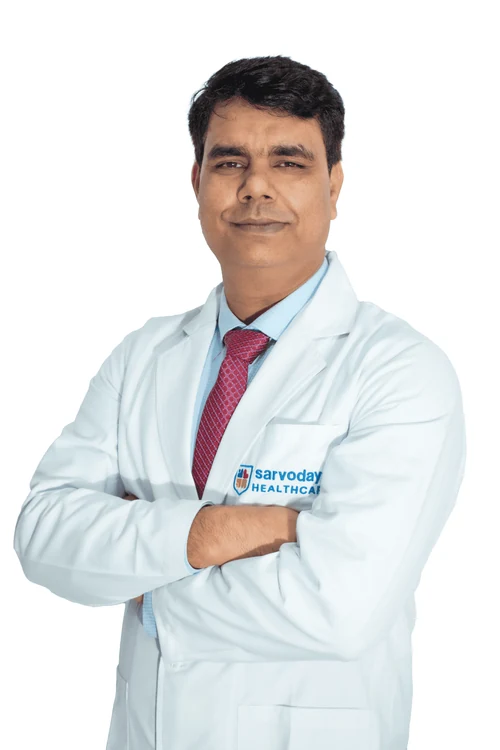
Why consult Dr. Naresh Panwar
- Cerebrovascular Neurosurgery is named among his specialisations on the Sarvodaya Hospital profile
- Brain haemorrhage surgery and brain stroke surgery are part of his listed areas of work
- MCh in Neurosurgery, SMS Medical College, Jaipur; earlier at Medanta Institute of Neurosciences, Lucknow
- Senior Consultant & Head Unit (II) – Neurosurgery at Sarvodaya Hospital, Sector 8, Faridabad
Questions to ask your surgeon
Write these down and bring them to your appointment.
- Does my lesion need treatment now, or can it be safely watched?
- What is my risk of bleeding if we do nothing?
- Is catheter treatment, surgery or radiation more suitable for me?
- Will I need seizure medicines afterwards?
- How often will I need follow-up scans?
Frequently asked questions
Does a neurosurgeon treat carotid artery narrowing?
Carotid narrowing is usually managed first with medicines and lifestyle change. If a procedure is considered, the choice is made by the vascular and neurology team after imaging.
Is every brain AVM or cavernoma operated on?
No. Many are monitored, especially if small, deep or never bled. The decision weighs bleeding risk against treatment risk for each person.
Kya dimag ki nas phatne ke baad hamesha operation hota hai?
Nahi. Kai mareezon ka ilaj ICU monitoring, BP control aur dawaiyon se hota hai. Operation tab socha jata hai jab khoon ka thakka dimag par dabav daal raha ho.
I live in NIT Faridabad. Where can I get my angiogram reviewed?
Bring your films and reports to Sarvodaya Hospital, Sector 8, Faridabad. Call +91 96608 88640 to confirm consultation timings.
Can I live normally with an untreated cavernoma?
Many people do, with blood pressure control, periodic MRI scans and prompt reporting of new symptoms.
Related care & reading
Appointments
Your brain and spine health deserve a carefully considered plan.
Request a consultation with Dr. Naresh Panwar at Sarvodaya Hospital, Sector 8, Faridabad. Bring your MRI or CT scans and reports, and the care team will help you prepare.