Skull Base · Complex Tumours
Skull Base Surgery in Faridabad
Lesions at the base of the skull sit among the nerves of sight, hearing, swallowing and facial movement. Protecting those functions is as important as removing disease.
खोपड़ी के निचले हिस्से (स्कल बेस) के ट्यूमर आँख, कान, चेहरे और निगलने की नसों के पास होते हैं। डॉ. नरेश पंवार की सूचीबद्ध विशेषज्ञता में स्कल बेस सर्जरी शामिल है, और हर मरीज़ के लिए रास्ता ट्यूमर की जगह के अनुसार चुना जाता है।
Understanding Skull Base Surgery
The skull base is the floor of the skull, the bony shelf on which the brain rests. Through small openings in it pass the nerves for smell, vision, eye movement, facial feeling and movement, hearing, balance and swallowing, along with the brain's major arteries and veins. Lesions here are often slow-growing, but space is so tight that even a modest tumour can affect these functions.
Skull base surgery is a listed specialisation for Dr. Naresh Panwar. Conditions in this region include acoustic neuroma (vestibular schwannoma), skull base meningiomas, pituitary tumours, craniopharyngioma, epidermoid cysts, clival lesions such as chordoma, and cerebrospinal fluid (CSF) leaks. Each needs a different route and a different balance of goals.
Depending on where a lesion sits, surgeons may use the endoscope through the nose, small 'keyhole' openings behind the ear or above the eyebrow, or conventional microsurgical approaches. For people in Faridabad, Palwal, Ballabhgarh and wider Delhi NCR, the first clue often comes from an ENT or eye check-up for hearing or vision change.
Common symptoms
Symptoms vary from person to person. Having one of these does not mean you have this condition.
A classic early sign of acoustic neuroma, often blamed on ear wax or age.
Unsteadiness, especially when walking in the dark.
From pressure on the trigeminal or facial nerves.
Involvement of the nerves that move the eyes.
Hoarseness or choking on liquids from lower cranial nerve involvement.
A constant watery drip, worse on bending, can be a CSF leak.
Possible causes & risk factors
Schwannomas grow from the covering of nerves such as the balance nerve.
Growing from the brain covering along the skull floor.
Craniopharyngioma and epidermoid cysts arise from cells left behind during development.
Chordoma and chondrosarcoma can arise from the clivus and nearby bones.
Can cause acoustic neuromas on both sides.
When to seek help
Book a routine consultation
- Hearing loss in one ear, especially with ringing or imbalance
- An MRI report mentioning a CP angle, clival or skull base lesion
- Gradual facial numbness, double vision or voice change
- Advice on watching versus treating an acoustic neuroma
Seek emergency care
- Sudden severe headache, vomiting or drowsiness
- Fever, neck stiffness and confusion in someone with a watery nasal drip (possible meningitis)
- Sudden trouble swallowing or breathing, or sudden vision loss: go to the nearest emergency department or call 112
How it is diagnosed
The central test, showing the lesion and its relation to nerves and vessels.
Shows bone erosion and the bony corridors used in surgery.
Measure hearing and help judge whether preserving it is realistic.
Maps arteries and veins close to the lesion.
Available treatment options
Treatment is chosen after examining you and studying your scans. Many people do well without an operation.
Small acoustic neuromas and other slow lesions are often watched with periodic MRI, as many grow very slowly or not at all.
Reaches midline lesions such as clival tumours and some meningiomas through the nose, using neuroendoscopy.
Approaches behind the ear (retrosigmoid) or at the side of the skull are used for lesions like acoustic neuroma.
Small acoustic neuromas or leftover tumour may be treated with highly focused radiation through the radiation oncology team.
Leaks through the skull base are sealed, often endoscopically, to lower the risk of meningitis.
When neurosurgery may be recommended
- A lesion growing on serial MRI
- Pressure on the brainstem or blocked brain fluid
- Progressive loss of vision or nerve function
- A persistent CSF leak, or a leak with past meningitis
- Tissue needed for diagnosis, as in a suspected chordoma
Procedure overview
A simplified outline. The exact steps depend on your scans and the approach chosen.
- Choosing the corridorThe route is chosen to reach the lesion while disturbing the fewest nerves and vessels.
- Guidance and monitoringImage guidance aids orientation, and nerve monitoring may help protect facial and other nerves.
- RemovalThe lesion is gently separated from nerves under high magnification.
- ReconstructionThe skull base is rebuilt with grafts or tissue flaps to prevent fluid leakage.
Potential benefits, risks & limitations
Potential benefits
- Relief of pressure on the brainstem and nerves
- Hearing, vision or facial function may stabilise or improve in selected cases
- Tissue diagnosis to guide further treatment
- Sealing a CSF leak lowers meningitis risk
Possible risks
- Facial weakness or numbness, temporary or lasting
- Hearing loss on the side of an acoustic neuroma
- Double vision, swallowing or voice difficulty
- CSF leak or meningitis
- Stroke or bleeding from injury to nearby vessels
When a tumour is stuck to vital nerves or arteries, a planned partial removal followed by observation or radiation is sometimes safer than chasing every last cell.
Recovery & follow-up
Recovery varies more than in most brain operations. Some patients go home within days after an endoscopic procedure; others need a longer stay with balance physiotherapy or speech-and-swallow therapy.
Eye care matters if facial weakness affects eyelid closure. Many skull base lesions need long-term MRI follow-up even after successful surgery.
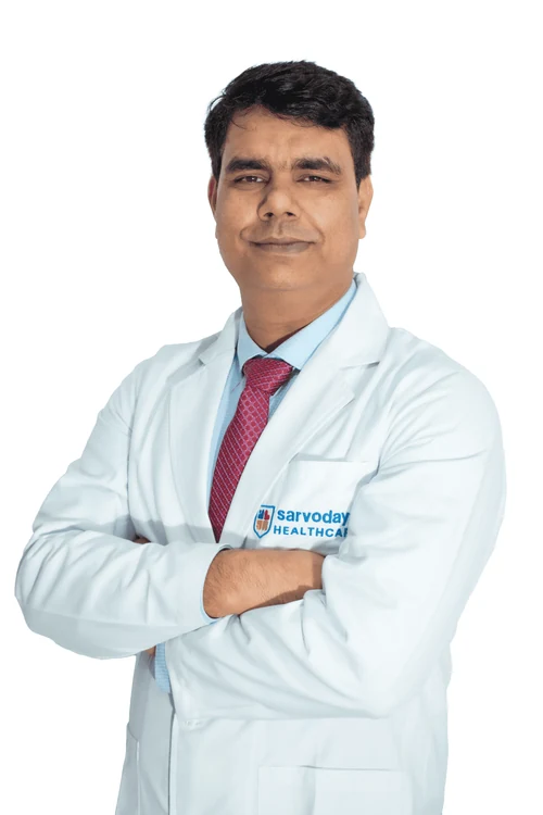
Why consult Dr. Naresh Panwar
- Skull Base Surgery is a listed specialisation on his Sarvodaya Hospital profile
- Fellowship in Neuroendoscopy, relevant to endoscopic skull base approaches
- Published case imaging includes meningioma cases
- Member of NSI and the European Association of Neurosurgical Societies (EANS)
Questions to ask your surgeon
Write these down and bring them to your appointment.
- Which approach do you suggest for my lesion, and why?
- What are the chances of keeping my hearing?
- Which nerves are at risk, and will nerve monitoring be used?
- Is observation or focused radiation an alternative?
- How long will I need follow-up MRI?
Frequently asked questions
What is an acoustic neuroma?
Also called vestibular schwannoma, it is a benign tumour on the balance nerve, beside the hearing nerve. It typically causes one-sided hearing loss, ringing and imbalance. Many small ones are watched; growing ones may need surgery or focused radiation.
What is a clival lesion?
The clivus is the sloping bone at the centre of the skull base, in front of the brainstem. Lesions here include chordoma and meningioma, and because they are deep they are often approached through the nose.
Skull base surgery ke baad chehra tedha ho sakta hai kya?
Kuch tumours chehre ki nas (facial nerve) ke bahut paas hote hain, isliye kamzori ka risk hota hai, jo temporary ya lambe samay tak ho sakti hai. Surgery se pehle doctor aapke tumour ke hisaab se ye risk samjhaate hain.
I have a watery drip from one nostril. Could it be serious?
Usually it is allergy, but a constant clear drip from one side, especially on bending forward, can be a CSF leak. It needs evaluation because it can lead to meningitis.
Is skull base surgery available in Faridabad?
Dr. Naresh Panwar consults at Sarvodaya Hospital, Sector 8, Faridabad, near Escorts Mujesar Metro Station. Call +91 96608 88640 to confirm consultation timings, and bring MRI, CT and hearing test reports.
Related care & reading
Appointments
Your brain and spine health deserve a carefully considered plan.
Request a consultation with Dr. Naresh Panwar at Sarvodaya Hospital, Sector 8, Faridabad. Bring your MRI or CT scans and reports, and the care team will help you prepare.