Brain Tumours · Meningioma
Meningioma Treatment in Faridabad
Most meningiomas are slow-growing and benign. The real question is timing: whether to watch, operate or use radiation, decided around you and not the scan alone.
मेनिन्जियोमा ज़्यादातर धीरे बढ़ने वाला, कैंसर-रहित ट्यूमर है। कई मरीज़ों में केवल नियमित MRI से निगरानी काफ़ी होती है, जबकि लक्षण या बढ़त होने पर सर्जरी या रेडियोथेरेपी पर विचार किया जाता है।
Understanding Meningioma
A meningioma grows from the meninges, the thin protective layers that wrap the brain and spinal cord. Because it grows from this covering rather than from brain tissue, it usually pushes the brain aside instead of invading it. Most meningiomas are WHO grade 1, benign and slow over years. A smaller number are atypical (grade 2) or, rarely, anaplastic (grade 3) and behave more actively.
Many meningiomas are found by chance, on an MRI done for headache or dizziness. Finding one does not automatically mean an operation. The choice between observation, surgery and radiotherapy depends on size, location, growth, symptoms, age and general health. Some sit deep at the skull base near nerves of vision and facial movement, where skull base surgery planning matters.
Surgeons describe how completely a meningioma was removed using Simpson grades. In simple words: Grade I means the tumour, its attachment to the brain covering and any affected bone were all removed. Grade II means the tumour was removed and its attachment sealed with heat. Grade III means the tumour was removed but the attachment left. Grade IV means part was deliberately left for safety, and Grade V means only a biopsy. Lower grades usually mean less chance of regrowth.
Common symptoms
Symptoms vary from person to person. Having one of these does not mean you have this condition.
Many small meningiomas are discovered incidentally and cause no trouble.
A dull headache whose pattern gradually changes over months.
Tumours on the brain's surface can irritate it and cause fits.
Tumours near the optic nerve may slowly dim vision in one eye.
Tumours under the frontal lobes can cause poor judgement or reduced smell, easily mistaken for ageing.
Possible causes & risk factors
Meningiomas are commoner in women, possibly linked to hormone receptors on tumour cells.
They become more frequent from middle age onwards.
Radiotherapy to the head, especially in childhood, raises the risk years later.
This inherited condition can cause several meningiomas.
When to seek help
Book a routine consultation
- A meningioma mentioned on an MRI done for another reason
- Gradual change in vision, smell, memory or behaviour
- Follow-up MRI shows the tumour has grown
- A second opinion before surgery or radiotherapy
Seek emergency care
- A first seizure or a prolonged fit
- Sudden worsening headache with vomiting or drowsiness
- Sudden vision loss or rapidly increasing weakness: go to the nearest emergency department or call 112
How it is diagnosed
Shows the tumour, its 'dural tail' attachment and any swelling in the brain next to it.
Shows calcium in the tumour and thickening of nearby skull bone.
Comparing scans months apart shows whether, and how fast, the tumour grows.
Important for tumours near the optic nerves.
Available treatment options
Treatment is chosen after examining you and studying your scans. Many people do well without an operation.
A repeat MRI, usually a few months to a year later, then longer intervals if stable. Many small tumours stay unchanged for years.
Anti-seizure medicines and short steroid courses may ease symptoms before or instead of surgery.
A craniotomy aims to remove the tumour with its attachment. For grade 1 tumours, complete removal often gives long-term control. See brain tumour surgery.
Focused radiation may suit small tumours in difficult spots, leftover tumour, or grade 2–3 tumours, planned with the radiation oncology team.
For tumours wrapped around vital nerves or vessels, safe partial removal followed by radiation or observation may protect function.
When neurosurgery may be recommended
- Symptoms such as seizures, weakness or vision loss
- Clear growth on repeat MRI
- Significant swelling in the brain around the tumour
- A large tumour pressing on the brain at diagnosis
- Scan features suggesting a higher grade
Procedure overview
A simplified outline. The exact steps depend on your scans and the approach chosen.
- MappingNavigation planning identifies nearby veins, nerves and vessels.
- Controlling blood supplyFeeding vessels at the tumour's base are controlled early to reduce bleeding.
- DebulkingThe centre is hollowed out so the edges can be gently peeled from the brain.
- Treating the attachmentThe dural base and involved bone are removed or sealed where safe, then the covering is repaired.
Potential benefits, risks & limitations
Potential benefits
- Complete removal of a benign meningioma often gives long-term control
- Pressure symptoms such as headache or weakness may improve
- Seizure control may improve
- Tissue diagnosis confirms the grade and guides follow-up
Possible risks
- Bleeding, especially in tumours with a rich blood supply
- Seizures or brain swelling after surgery
- Injury to nearby nerves, vessels or venous sinuses
- Infection or cerebrospinal fluid leak
- Recurrence, even after complete removal
When a meningioma is wrapped around critical nerves or arteries, removing every last piece may carry too high a risk, so a small residue may be left and watched or treated with radiation.
Recovery & follow-up
Recovery depends greatly on location. Many patients with surface tumours walk the next day, while skull base operations may need a longer stay. Tiredness is common for some weeks.
Because meningiomas can return slowly, follow-up MRI continues for many years at widening intervals. Driving and return to work are discussed individually, especially if seizures occurred.
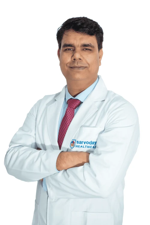
Why consult Dr. Naresh Panwar
- Published case imaging includes meningioma cases
- Skull Base Surgery is a listed specialisation on his Sarvodaya Hospital profile
- MCh (Neurosurgery), SMS Medical College, Jaipur; MS (General Surgery), KGMU, Lucknow
- Previously Senior Consultant, Neurosurgery at Medanta Institute of Neurosciences, Lucknow
Questions to ask your surgeon
Write these down and bring them to your appointment.
- Is my meningioma growing, and how fast?
- Is it safe to wait and repeat the MRI?
- What Simpson grade of removal do you expect, and why?
- Which nerves or vessels are close to the tumour?
- Would radiotherapy be a reasonable alternative?
Frequently asked questions
My MRI shows a small meningioma but I feel fine. Do I need surgery?
Often not. Many small meningiomas without symptoms or swelling are safely watched with repeat MRI. Surgery is considered if the tumour grows, causes symptoms or looks higher grade.
Can a meningioma come back after surgery?
Yes, even after complete removal, though the chance is generally lower after Simpson grade I–II removal. Grade 2 and 3 tumours recur more often, so regular follow-up MRI matters.
Kya meningioma cancer hota hai?
Zyadatar meningioma cancer nahi hote; ye dheere badhne wale benign tumour hote hain. Kuch kam cases mein ye atypical ya aggressive ho sakte hain, isliye operation ke baad biopsy report zaroori hai.
Is radiotherapy an option instead of surgery?
For some small tumours in risky locations, focused radiation may be considered. It aims to stop growth rather than remove the tumour. The radiation oncology team assesses suitability.
I am from Palwal. Can I get my meningioma reports reviewed?
Yes. Consultations are at Sarvodaya Hospital, Sector 8, Faridabad. Please call +91 96608 88640 to confirm timings and Palwal consultation availability, and bring previous MRI scans on CD.
Related care & reading
Appointments
Your brain and spine health deserve a carefully considered plan.
Request a consultation with Dr. Naresh Panwar at Sarvodaya Hospital, Sector 8, Faridabad. Bring your MRI or CT scans and reports, and the care team will help you prepare.