Brain · Keyhole Surgery
Neuroendoscopy in Faridabad
A slim camera can let surgeons reach deep parts of the brain through a small opening or through the nose — when the condition and anatomy are right for it.
न्यूरोएंडोस्कोपी में पतले कैमरे की मदद से छोटे छेद या नाक के रास्ते दिमाग के अंदर ऑपरेशन किया जाता है। यह हर मरीज़ के लिए सही नहीं होता — फैसला जांच और स्कैन के आधार पर होता है।
Understanding Neuroendoscopy
A neuroendoscope is a slim tube carrying a high-definition camera and light. Through it, or alongside it, the surgeon passes fine instruments to work inside the brain's fluid spaces or at the skull base. Because the camera brings the view close to the target, the opening can be small — a single burr hole, or no external cut at all when the route is through the nostril.
Neuroendoscopy is a technique, not a disease. It is used for blocked fluid pathways (hydrocephalus), fluid-filled cysts, deep tumours that need a tissue sample, and tumours of the pituitary gland and skull base. The same camera-guided principle is also used in the spine.
Dr. Naresh Panwar holds a Fellowship in Neuroendoscopy, and neuro-endoscopy is listed among his specialisations at Sarvodaya Hospital, Sector 8, Faridabad. One honest point matters: smaller is not automatically safer. For some tumours, a conventional microscope operation gives a clearer view or more complete removal, and that choice is explained case by case.
Common symptoms
Symptoms vary from person to person. Having one of these does not mean you have this condition.
May indicate blocked fluid flow from a cyst or narrowing.
A pituitary tumour pressing on the optic nerves can narrow vision at the edges.
Irregular periods, unexpected breast milk or enlarging hands and feet may point to the pituitary.
Can result from fluid build-up or a cyst pressing on brain pathways.
A persistent salty, clear drip from one nostril can be a CSF leak at the skull base.
Possible causes & risk factors
A blockage in the fluid channels, often suited to endoscopic third ventriculostomy.
Fluid-filled sacs that can press on brain or block drainage.
Mostly non-cancerous growths, often reachable through the nose.
An endoscopic biopsy may give a diagnosis through one small opening.
Defects letting fluid leak through the nose, sometimes repairable endoscopically.
When to seek help
Book a routine consultation
- A cyst or ventricle enlargement noticed on MRI
- A pituitary tumour diagnosed by an endocrinologist or eye specialist
- A second opinion on whether keyhole surgery suits your case
Seek emergency care
- Sudden severe headache with drop in vision or double vision — go to the nearest emergency department or call 112
- Repeated vomiting, drowsiness or confusion
- Fever with neck stiffness, especially with watery nasal discharge
- A seizure or sudden weakness
How it is diagnosed
The key planning scan — shows the lesion and the fluid pathways the endoscope will travel through.
Maps bone and sinus anatomy before surgery through the nose.
Essential for pituitary tumours, usually coordinated with endocrinology and ophthalmology.
Image guidance can plan a precise path to deep targets.
Available treatment options
Treatment is chosen after examining you and studying your scans. Many people do well without an operation.
Many small cysts and some pituitary tumours are simply followed with scans.
Prolactin-secreting tumours often shrink with medicine alone, so surgery may not be needed.
A new drainage route can be made for blocked fluid, a cyst wall opened so it drains, or a small tissue sample taken from a deep tumour.
Pituitary and selected skull base tumours are reached through the nostril without a scalp incision.
Camera-guided techniques also treat some disc and nerve problems — see endoscopic spine surgery.
When neurosurgery may be recommended
- Symptoms clearly linked to a cyst or fluid blockage
- A pituitary tumour threatening vision or not controlled by medicines
- A deep lesion needing diagnosis before further treatment
- MRI anatomy that allows a safe endoscopic route
Procedure overview
A simplified outline. The exact steps depend on your scans and the approach chosen.
- Planning the corridorA burr hole is chosen for ventricular work, the nostril for skull base work.
- General anaesthesiaThe patient is fully asleep; navigation may be linked to the scans.
- Camera-guided surgeryThe surgeon works from a magnified video image with fine instruments.
- Closure and early checksEndonasal openings may be sealed with the patient's own tissue; hormone, fluid and neurological checks follow.
Potential benefits, risks & limitations
Potential benefits
- Smaller openings and often less disturbance to normal brain
- No visible scalp incision for many endonasal operations
- May avoid a shunt in suitable hydrocephalus
- Often a shorter hospital stay, though this varies
Possible risks
- Bleeding, which can be harder to control through a narrow channel
- Infection, including meningitis
- CSF leak through the nose after endonasal surgery
- Hormone imbalance or excess urination after pituitary surgery
- Need to switch to open surgery during the operation
Endoscopy cannot safely remove every tumour, especially large, very vascular or widely spread ones. A keyhole operation is still brain surgery with real risks.
Recovery & follow-up
After a ventricular procedure, many patients are walking within a day or two. After surgery through the nose, stuffiness and crusting are common for some weeks; avoiding nose-blowing and straining is usually advised.
Pituitary patients need hormone follow-up, sometimes long term. Patients coming from Palwal or Greater Faridabad can plan wound checks and reviews in advance; please call to confirm consultation timings.
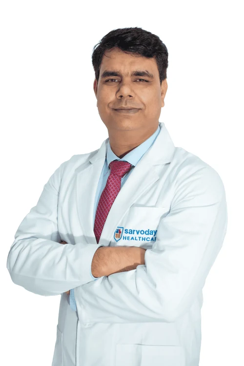
Why consult Dr. Naresh Panwar
- Completed a Fellowship in Neuroendoscopy in addition to his MCh in Neurosurgery
- Neuro-endoscopy and skull base surgery both appear on his Sarvodaya Hospital specialisation list
- His published case imaging includes a navigated (frameless) brain biopsy
- Head Unit (II) – Neurosurgery at Sarvodaya Hospital, with more than a decade of neurosurgical practice
Questions to ask your surgeon
Write these down and bring them to your appointment.
- Is an endoscopic approach realistic for my condition, or is open surgery safer?
- What happens if the operation cannot be completed endoscopically?
- Will I need hormone tests or an endocrinologist afterwards?
- How should I care for my nose after endonasal surgery?
- What is the chance the cyst or tumour comes back?
Frequently asked questions
Is neuroendoscopy the same as laser brain surgery?
No. Neuroendoscopy uses a camera and fine instruments, not a laser beam. Keyhole describes the small opening, not a different kind of energy.
Kya naak ke raste brain tumour nikala ja sakta hai?
Kuch tumours, khaaskar pituitary tumours, endoscope ki madad se naak ke raste nikale ja sakte hain. Lekin har tumour is tareeke ke layak nahi hota — MRI aur jaanch ke baad hi tay hota hai.
Will there be a visible scar?
Ventricular endoscopy usually leaves a small scar hidden in the hair. Endonasal surgery usually leaves no visible scar on the face.
Is endoscopic surgery always preferable to microscope surgery?
No. Each has strengths for different lesions. Our article on what neuroendoscopy is explains the differences.
Can I get a neuroendoscopy opinion in Faridabad?
Yes. Consultations are at Sarvodaya Hospital, Sector 8, Faridabad, near Escorts Mujesar metro station on the Violet Line. Call +91 96608 88640 to confirm timings and bring all your scans.
Related care & reading
Appointments
Your brain and spine health deserve a carefully considered plan.
Request a consultation with Dr. Naresh Panwar at Sarvodaya Hospital, Sector 8, Faridabad. Bring your MRI or CT scans and reports, and the care team will help you prepare.